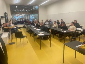
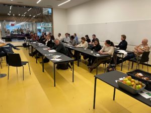
On 24 and 25 of November in DTU was held DanScatt X-Ray Bioimaging Workshop!
It contained a lot of interesting lectures, introductions of different X-Ray and Bioimaging centres and discussions.
Part of the Workshop topics were:
- Science highlights:
- Nina K. Wittig, a facility manager in AXIA – the Aarhus X-ray Imaging Alliance, talked about Multiscale X-ray computed tomography in bone research or how they in AXIA characterize complex materials such as bone from the nano- to the organ-scale using a combination of X-ray CT equipment ranging clinical CT to laboratory instrumentation to advanced synchrotron imaging
- Jessica Pingel from Department of Neuroscience of University of Copenhagen presented Synchrotron imaging of muscle tissue – shedding light on muscle atrophy in neuromuscular disorders
- Paola Coan, an Associate Professor at the Ludwig Maximilian University (Faculties of Medicine & Physics) made presentation on Coherent Synchotron X-rays to Elucidate Anatomy Pathology and Therapy and key requirements of biomedical imaging as well as the main challenges in the field have been discussed
- Eva Christensen, from Department of Green Technology of University of Southern Denmark talked about Imaging proteins in cells using super-resolved microscopy and advanced analysis tools.
- Imaging at various research facilities:
- Alberto Astolfo, System Engineer at the Advanced X-Ray Imaging Group in University College London showed An overview of the activities at the UCL Advanced X-Ray Imaging Labs, where he reviewed the main imaging methods that have been developed so far and discussed their applications across the life and physical sciences.
- Clara Prats, part of Core facility for Integrated Microscopy of University of Copenhagen made introduction to Danish Bioimaging Network
- Jonathan Brewer, from the Danish Molecular Bioimaging Center of University of Southern Denmark presented the Applications of Advanced Optical Microscopy at the Danish Molecular Biomedical Imaging Center (DaMBIC), where he talked about some of the imaging techniques including Coherent anti stokes Raman scattering microscopy, which can deliver chemically specific imaging at video rate, and the new MINFLUX microscope in DaMBIC.
- Mathilde Lerche from DTU Health introduced the facility of Multimodal and Translational Bio-Imaging at DTU and provided examples of on-going research
- Karina Thånell, a Beamline Scientist at the SoftiMAX beamline at MAX IV and a Group Manager of the Imaging Beamlines group introduced the Bio-Imaging opportunities at MAX IV
- Science talk:
- Alexander Schulz, from the Department of Plant & Environment Sciences, of University of Copenhagen talked about Breaking the limits of live vascular imaging in plants by µXCT
- Søren Husted, professor at University of Copenhagen presented the collaboration between KU, DTU and McGill University (Canada) on Biocompatible nano fertilizers for targeted delivery and programmed release of essential nutrients in crops – Imaging of basic plant nanoparticle interactions, which is a visionary approach that bypasses nutrient fixation and immobilization in the soil.
- Image analysis challenges:
- Anders Dahl, professor at DTU Compute made an introduction of QIM – the Center for Quantification of Imaging Data from MAX IV
- Florian Schott, PhD student from Lund University presented the Foamquant- 4D image quantification toolbox for cellular materials
- Jon Sporring from Department of Computer Science of University of Copenhagen talked about 3D image quantification cases
We want to thank to all participants and invited lecturers for being part of the workshop!